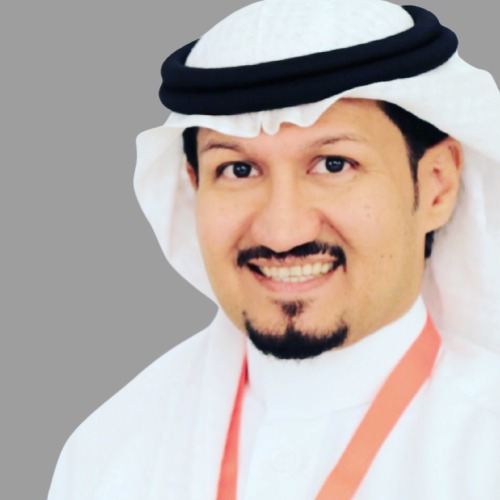
Dr. Hassan Koshak
Ministry of Interior Security Forces Medical Services
Hassan Koshak has been a consultant in periodontics and implant dentistry and has been the head of the dental department at Comprehensive Specialized Polyclinic, Ministry of Interior Security Forces Medical Services, Jeddah, KSA, since 2016. He holds a Saudi Fellowship in Dental Implant (2014-2016) and a Saudi Board in Periodontics (2012-2014) from the Saudi Commission for Health Specialties. He earned his MSD and Clinical Certificate in Periodontics (with honors) from Riyadh Colleges of Dentistry and Pharmacy (2009-2012), Advanced Education in General Dentistry (AEGD) from the University of Southern California (2006-2008), and BDS from King Abdul-Aziz University, Jeddah, KSA.
Saudi Arabia
Abstracts
Short implant Vs sinus elevation
Clinical choice of the most appropriate implant therapy modality should be based on an assessment of the residual alveolar bone height, width, and sinus morphology with a cone beam computed tomography (CBCT) scan, current scientific evidence, surgical skills and experience of the surgeon, and the patient’s preferences.
Following a good surgical protocol and an excellent oral hygiene maintenance program are fundamental elements in achieving a successful and predictable outcome.
The available evidence on short dental implants in early research was not significant compared with the longer dental implants; the surface treatment is improving now than before for this reason.
The use of short implants allows the treatment of patients who are unable to undergo complex surgical techniques for medical, anatomic, or financial reasons. By reducing the need for complex surgeries short implants reduce morbidity, cost, and treatment time. Recently short implants offer a less invasive treatment alternative in resorbed ridge cases.
Can alveolar ridge be completely preserved by partial extraction therapies (PET): A case Report
Introduction:
Tooth extraction is usually followed by partial resorption of the residual alveolar ridge. Different techniques such as the ridge preservation procedure have been proposed to maintain the ridge dimension. However, applying these methods to extraction sockets could not completely preserve the coronal part of facial bone walls which were comprised almost entirely of bundle bone.
Aim:
To assess the effectiveness of partial extraction therapy (PET) for complete preservation of alveolar ridge.
Case report:
A 38-year-old woman with non-contributory medical history presented to our clinic with non-restorable tooth #12, which doesn’t have any periapical or periodontal pathology. After clinical and radiographically assessment, computed tomography (CBCT), indicated insufficient width of buccal bone plate therefore partial extraction therapies were planned for simultaneous immediate implant placement (Nobel Biocare 3.5x13mm Conical implant with Cover screw) and provisionalization by Acrylic restoration. Initial follow up after two weeks, then after 2 months final restoration by screw retained crown inserted. After 6 months, 1 and 3 years of loading follow up by radiographical assessment, for evaluation of bone remodeling and clinical evaluation of soft tissue changes around the implant.
Results:
Two weeks follow up revealed the healing was uneventful, and after 6 months, 1 and 3 years the clinical and radiographical assessment revealed, that retaining root fragment adjacent to the buccal crestal bone and placing an implant engaged to the palatal socket wall immediately are able to maintain the contour of the ridge. The implant can achieve osseointegration without any inflammation at peri-implant tissue and also soft tissue contour preserved.
Discussion:
PET is technique-sensitive and associated with the risk of displacement of the buccal root fragment, or even the buccal lamellar bone. The root fragment should be reduced along its vertical axis to the level of the height of the alveolar ridge to prevent perforation of the buccal mucosa during the healing period.
Encountered when managing the post-extraction tissues. This case report of immediate implant placement simultaneous to the PET is among the first to demonstrate with a 3 years follow up successful preservation of post-extraction tissues coinciding with successful restorative implant treatment.
Conclusion:
After three years follow up, PET can prevent soft and hard tissue changes that can happen during the healing of the alveolar socket after tooth extraction. However, the use of PET as routine clinical practice still needs to higher level of evidence.
Dental suturing materials and techniques
Successful dental suturing or oral surgery is dependent on accurate coaptation of the flaps. Various methods and materials have been used (sutures, stents, paste dressings, tissue tacks, and adhesives) for precise flap placement. Suturing has remained the most popular method.
The term “suture” describes any strand of material utilized to ligate blood vessels or approximate tissues.
The primary objective of dental suturing is to position and secure surgical flaps in order to promote optimal healing (first/primary intention) provide support for tissue margin until they heal, without dead space, and reduce postoperative pain.
Inadequate suturing may result in flap skipping, exposed bone/necrosis, pain, and delayed wound healing. A variety of suture materials and suture/needle combinations are available. The choice of suture for a particular procedure is based on the known physical and biologic characteristics of the suture material and the healing properties of the sutured tissues.
The selection of suture material is based on:
The condition of the wound, the tissues to be repaired, the tensile strength of the suture material, knot-holding characteristics of the suture material, and the reaction of surrounding tissues to the suture materials.
Knowledge of the suture, needles (type, size, shape), instruments, and techniques are absolutely necessary in order to be a competent surgeon.
There is no suture superior to the others in each aspect.
The differences in terms of tissue reaction and bacterial adhesion between sutures should be always considered in the selection of the appropriate suturing material.
Delicate and proper soft tissue handling during various suturing techniques can ensure optimal tissue healing and high esthetic results.
Featured 2025 Speakers
Speakers of The Club
Dr. Tarek Assi
Alpha Dental Practice & Implant Center
Dr. Abdalhadi Kawaiah
A Kawaiah Orthodontics
Dr. Ammaar Abidi
Lincoln Memorial University

Dr. Rusul Yaseen
Certified Smile Makeover

Dr. Bak Nguyen
CEO - Mdex & Co.
Dr. Boros Loredana
Dental One Studio

Dr. Oksana Soroka
CEO - GScompany_ed
Prof. Dr. Niladri Maiti
Central Asian University
Dr. Aiman Obeid
Specialist Orthodontist

Dr. Wahiba Lagmou
The Moroccan Academy of Endodontics
Dr. Beatriz Chaves
University of the Basque Country

Dr. Jasmine Haddad
Smart Dental Boss

Prof. Dr. Sherin Khalam
London Academy of Dentistry
Dr. Lina Maiza
University of Algiers
Dr. Yaniv Mayer
Rambam Medical Center
Prof. Dr. Preetinder Singh
Academy of Oral Surgery

Prof. Dr. Annamalai Thangavelu
Annamalai University
Dr. Jasdeep Kaur Cheema
Nuvia Dental Implant Center

Dr. Sara Massraf
The Great Western Hospital
Dr. Lubna Alolaiwi
Riyadh Elm University
Dr. Nosirdzhon Karimov
Sahih Digital Med
Prof. Dr. Robertha Tulio
President of CRORJ's Technical Chamber of Anesthesia and Sedation in Dentistry